Tuesday 4 October 2011

A 50 year old man with chest pain for 24 hours


Ventricular bigeminy

  • a ventricular premature beat follows each normal beat
There are also features of an acute inferior myocardial infarction.


Ventricular bigeminy

Long QT interval

  • The QT interval normally varies with heart rate - becoming shorter at faster rates. It is usually corrected using the cycle length (R-R interval) as shown opposite.
  • normal QTc = 0.42 seconds
Romano-Ward syndrome is an autosomal dominantly inherited form of long QT interval and there is a risk of recurrent ventricular tachycardia, particularly Torsade de Pointes.

Ventricular premature beats (VPBs)

  • 2 ventricular premature beats are also shown in this ECG
  • They are
    • broad
    • occur earlier than normal
    • and are followed by a full compensatory pause (the distance between the normal beats before and after the VPB is equal to twice the normal cycle length).

long qt interval

A 22 year old lady with prolonged vomiting.


This lady's serum potassium was 1.8 mmol/L.

Hypokalaemia

The following changes may be seen in hypokalaemia.

  • small or absent T waves
  • prominent U waves (see diagram)
  • first or second degree AV block
  • slight depression of the ST segment


hypokalemia


A 58 year old man on haemodialysis presents with profound weakness after a weekend fishing trip.


Hyperkalaemia

The following changes may be seen in hyperkalaemia

  • small or absent P waves
  • atrial fibrillation
  • wide QRS
  • shortened or absent ST segment
  • wide, tall and tented T waves
  • ventricular fibrillation

hyperkalemia

Monday 19 September 2011


Alex is a 4 weeks old baby. He was born at term, normal delivery with no complication. From 3 weeks old, he started to be irritable especially during breast feeding. Mum noticed that he was sweating during feeds, and seems to be distressed. His grandmother thinks that he has “reflux”.
Past few days the weather have been very cold. Alex was brought into Emergency Department with a low grade fever, rhinorrhea and poor feeding. On examination, he was found to be tachypnic and tachycardic. There was a soft systolic murmur. Few crepitation was heard over the lung field. Liver is palpable 3cm below the costal margin. An ECG was done as part of the work up for the murmur.
What is the most likely diagnosis.
A) Bronchiolitis
B) Tetralogy of Fallot
C) Anomalous origin of the left coronary artery arising from the pulmonary artery
D) Coarctation of the aorta
E) Ventricular Septal Defect

paediatric ecg

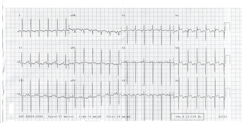
A 5 years old boy with Atrial Septal Defect attended for pre-operative medical examination. He had a irregular pulse. Attached is the ECG of this child.


1. What is the diagnosis?
A) Sinus arrhythmia
B) Wandering Atrial Pacemaker
C) Atrial Fibrilation
D) Complete Heart Block
E) Second Degree Heart Block
2. What is the significance of this finding?
A) no clinical significance
B) This child is at risk of sudden death from cardiac arrhythmias
C) This child is in cardiac failure
D) This child needs a permanent cardiac pacemaker implantation
E) This child needs anticoagulation

atrial septal defect

Sunday 18 September 2011


A 79 year old man with 5 hours of chest pain.

Acute myocardial infarction in the presence of left bundle branch block

Features suggesting acute MI

  • ST changes in the same direction as the QRS (as shown here)
  • ST elevation more than you'd expect from LBBB alone (e.g. > 5 mm in leads V1 - 3)
  • Q waves in two consecutive lateral leads (indicating anteroseptal MI)

Acute myocardial infarction in the presence of left bundle branch block

comments